Home

Hose Software Mannschaft imaris cell counting Zwei Grad Hilflosigkeit Besucher

Longitudinal Imaris cell counts in MDA-MB-231 aggregates before and... |  Download Scientific Diagram
Longitudinal Imaris cell counts in MDA-MB-231 aggregates before and... | Download Scientific Diagram
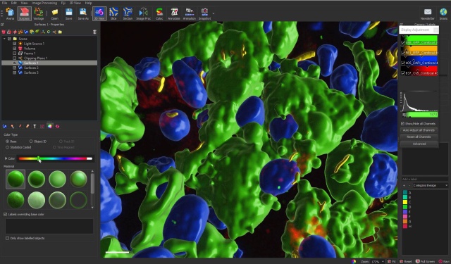
Imaris Part 4: Object Classification - ZMB UZH

Count Spots Inside the ROI - Tutorial- Oxford Instruments

Imaris 9.5 - 3D/4D Image Visualization & Analysis with Deconvolution |  Microscopy Sotware - Imaris - 牛津仪器
Imaris 9.5 - 3D/4D Image Visualization & Analysis with Deconvolution | Microscopy Sotware - Imaris - 牛津仪器
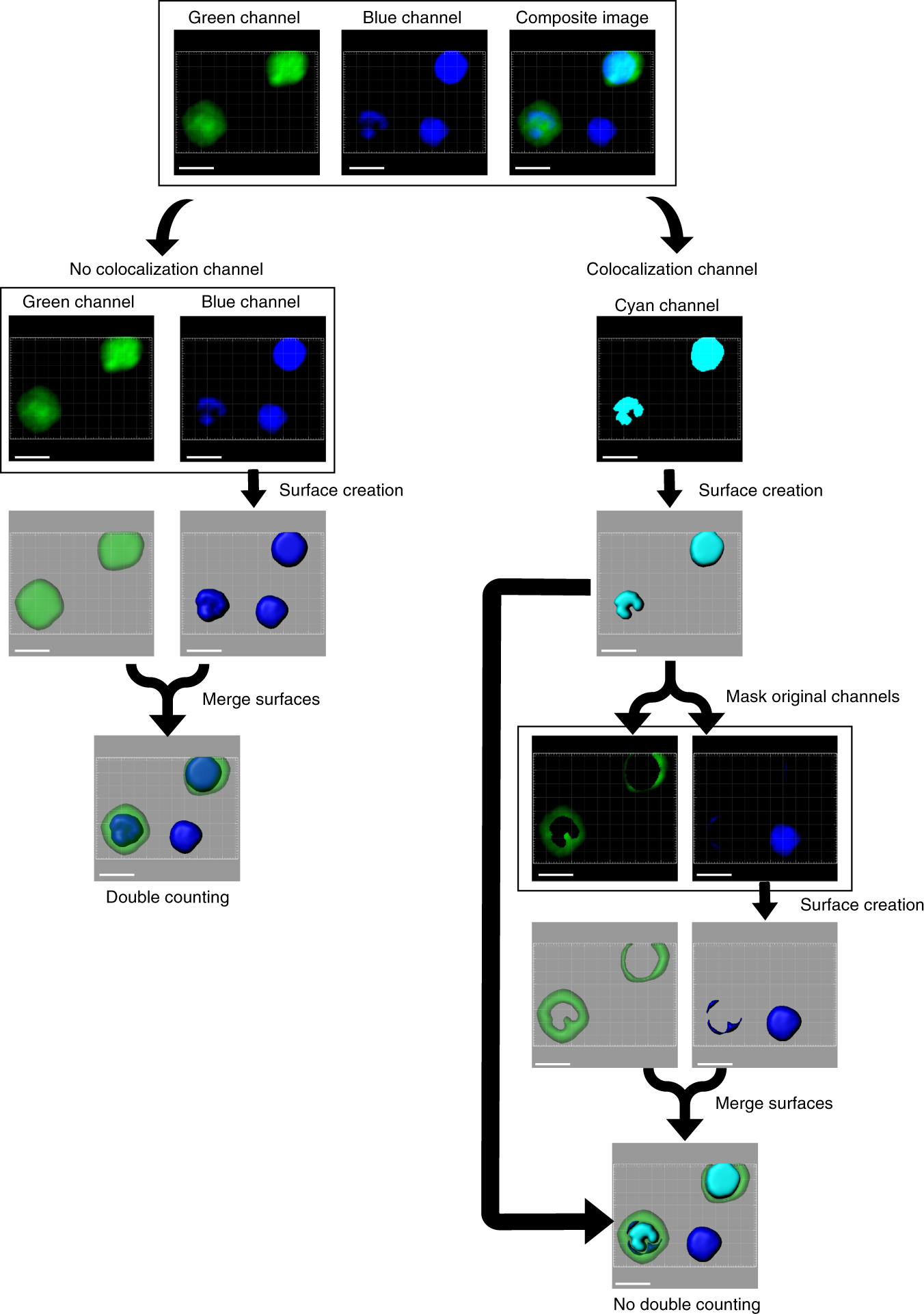
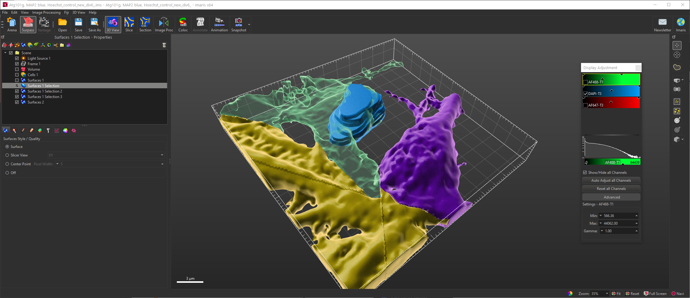
Streamlining volumetric multi-channel image cytometry using  hue-saturation-brightness-based surface creation | Communications Biology
Streamlining volumetric multi-channel image cytometry using hue-saturation-brightness-based surface creation | Communications Biology
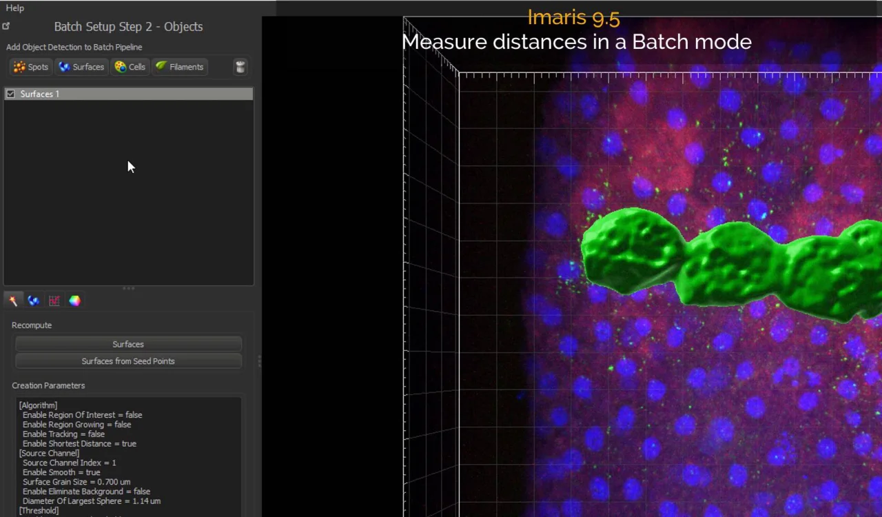
Any advice on automated neuron count in Imaris? | ResearchGate

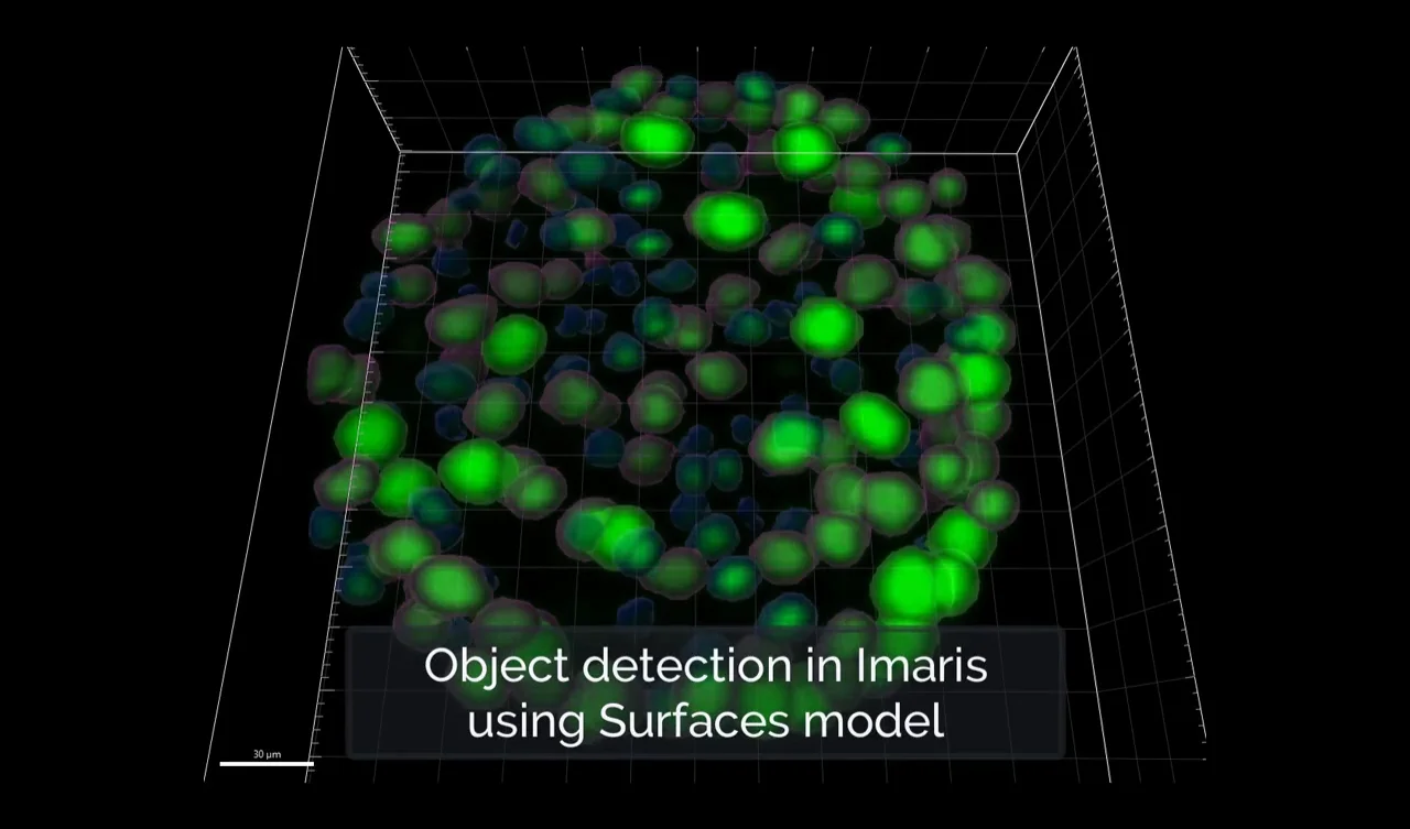
Learn how to detect objects in Imaris using the Surfaces model- Oxford  Instruments
Learn how to detect objects in Imaris using the Surfaces model- Oxford Instruments

Software – Cellular Imaging Facility

Imaris for Cell Biologists - Imaris - Oxford Instruments

Imaris for Neuroscientists - Imaris - Oxford Instruments

Imaris for Cell Biologists - Imaris - Oxford Instruments

Imaris 9.5 - 3D/4D Image Visualization & Analysis with Deconvolution |  Microscopy Software - Imaris - Oxford Instruments
Imaris 9.5 - 3D/4D Image Visualization & Analysis with Deconvolution | Microscopy Software - Imaris - Oxford Instruments
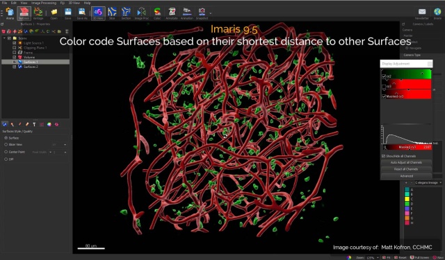
Automated Cell Counting in ImageJ - YouTube

Imaris 9.5 - 3D/4D Image Visualization & Analysis with Deconvolution |  Microscopy Sotware - Imaris - 牛津仪器
Imaris 9.5 - 3D/4D Image Visualization & Analysis with Deconvolution | Microscopy Sotware - Imaris - 牛津仪器

How to Perform Live/Dead Quantification Using Imaris - Allevi

Histological characterization and quantification of newborn cells in the  adult rodent brain: STAR Protocols
Histological characterization and quantification of newborn cells in the adult rodent brain: STAR Protocols
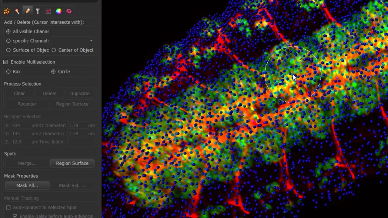
Imaris 9.2 - Count & track cells, nuclei & vesicles faster than ever before  - Imaris - Oxford Instruments
Imaris 9.2 - Count & track cells, nuclei & vesicles faster than ever before - Imaris - Oxford Instruments
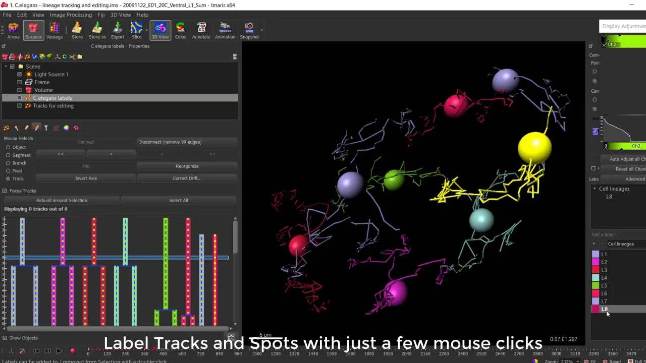
Imaris 9.2 - Count & track cells, nuclei & vesicles faster than ever before  - Imaris - Oxford Instruments
Imaris 9.2 - Count & track cells, nuclei & vesicles faster than ever before - Imaris - Oxford Instruments

Advanced Image Processing using Imaris (25. 11. 2008)

Imaris 9.2 - Count & track cells, nuclei & vesicles faster than ever before  - Imaris - Oxford Instruments
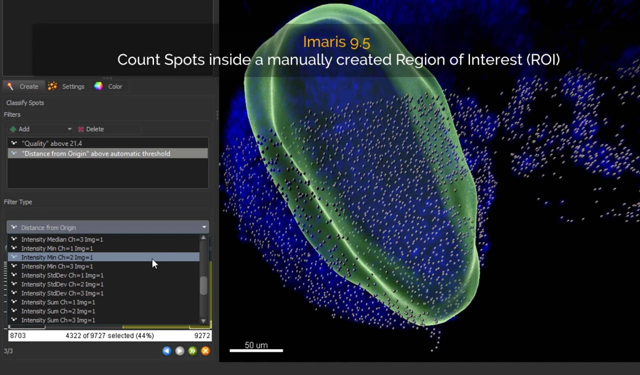
Imaris 9.2 - Count & track cells, nuclei & vesicles faster than ever before - Imaris - Oxford Instruments

Imaris for Core Facilities | Full Power of Imaris at your Fingertips -  Imaris - Oxford Instruments
Imaris for Core Facilities | Full Power of Imaris at your Fingertips - Imaris - Oxford Instruments
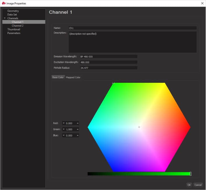
Imaris for Cell Biologists - Imaris - Oxford Instruments
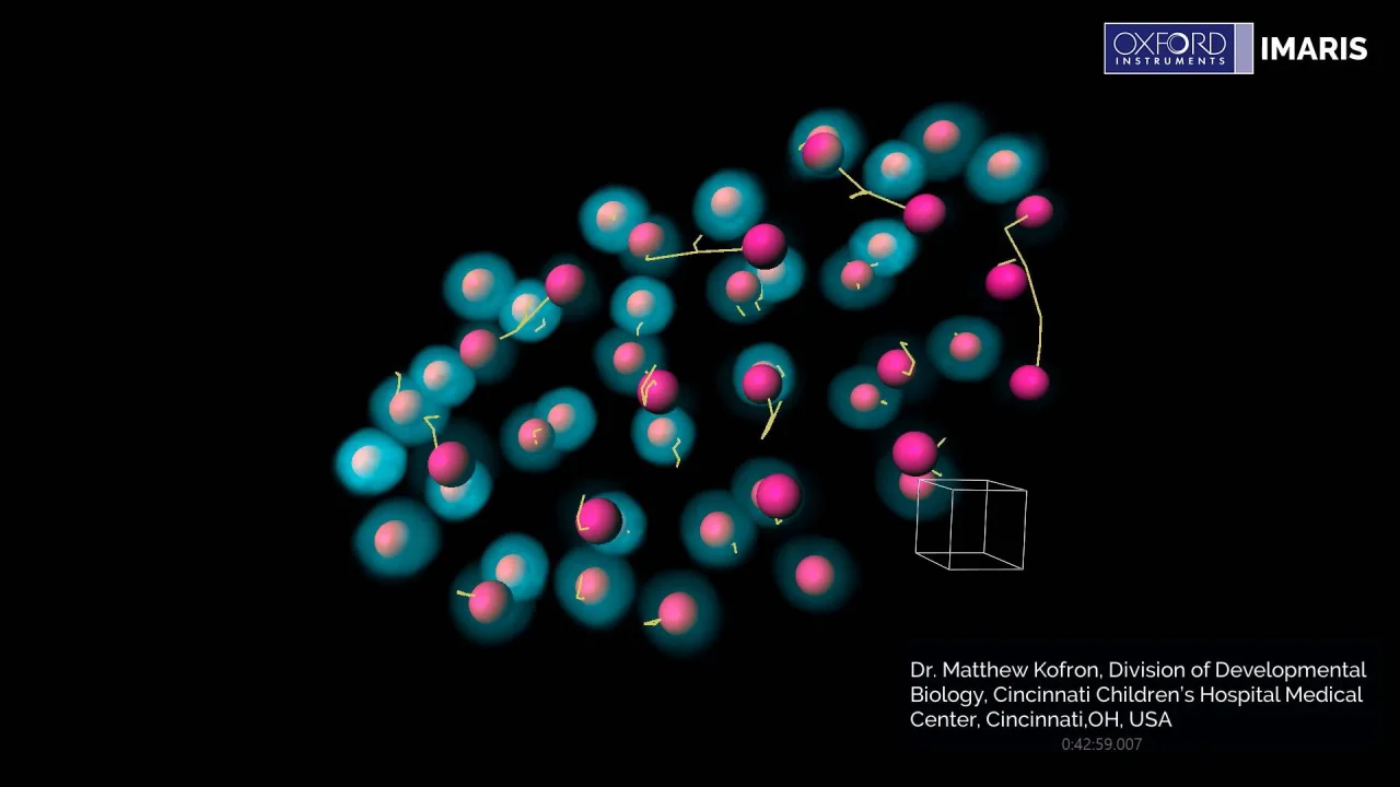
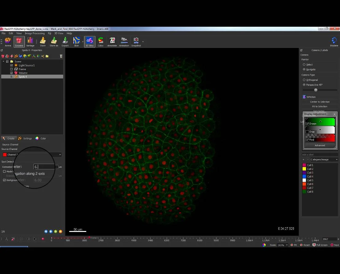
3D/4D Image Analysis for Cell Biologists in Imaris | Webinar- Oxford  Instruments
3D/4D Image Analysis for Cell Biologists in Imaris | Webinar- Oxford Instruments